
Knee Pain Location Chart: What Your Pain is Telling You
Knee pain is a near-universal experience. Whether it’s a dull ache after a long day, a sharp twinge when you stand up, or persistent discomfort that limits your life, millions of people search for answers every day. It’s one of the most common complaints that brings people to a specialist, affecting individuals of all ages and lifestyles, from athletes to office workers. But “knee pain” is a broad term. The key to finding relief lies in understanding where it hurts.
This is where a knee pain location chart becomes an invaluable tool. Think of it as a map to your discomfort. By pinpointing the exact area of your pain—be it the front, back, inner, or outer side—you can gain crucial insights into the potential underlying cause. This knowledge empowers you to have a more informed conversation with your doctor, leading to a faster, more accurate knee pain diagnosis and a more effective treatment plan.
In this comprehensive guide, we will walk you through the anatomy of the knee and explore a detailed knee pain chart. We will be guided by the expertise of Dr. Hesham Al Khateeb, an award-winning, UK Board Certified Hip and Knee Surgeon with a wealth of experience in treating complex knee conditions. With over 2,000 joint replacements performed, Dr. Al Khateeb’s insights will help demystify your symptoms and set you on the path to recovery.
First, Are You Experiencing These Telltale Signs?
Before we dive into the knee pain location chart, it’s important to recognize the common symptoms that accompany knee issues. While pain is the primary indicator, it often comes with friends. If you’re experiencing any of the following, your knees are trying to tell you something is wrong:
Joint Swelling and Stiffness: Is your knee puffy, swollen, or difficult to move, especially in the morning?
Pain When Bending or Straightening: Do simple movements like sitting down or extending your leg cause a sharp or aching pain? This is a classic sign that an internal structure is irritated.

Popping or Locking Sensations: A feeling of the knee “catching,” locking in place, or making grinding/popping sounds can indicate cartilage or meniscus issues.
Weakness or Instability: Do you feel like your knee might “give way” or buckle when you put weight on it?
Redness and Warmth: Skin over the joint that is red or warm to the touch can signal inflammation or infection.
Numbness or Tingling: A less common but important symptom that could indicate nerve involvement.
Recognizing these signs is the first step. The next is understanding the complex machinery within your knee that could be causing them.
A Simple Look at Your Knee's Anatomy
To understand a knee pain location chart, you first need a basic understanding of your knee’s structure. Your knee isn’t just a simple hinge; it’s a complex joint responsible for bearing your body’s weight and enabling movement. Dr. Hesham Al Khateeb emphasizes that understanding this knee anatomy pain connection is vital for patients.
Let’s break it down into four main components:
Bones
The knee joint is where three main bones meet:
Femur (Thighbone): The large bone at the top of the joint.
Tibia (Shinbone): The primary weight-bearing bone in your lower leg.
Patella (Kneecap): The small, flat bone that sits in the front, protecting the joint and providing leverage for your muscles. When you experience knee bone pain, it often involves the interaction between these surfaces.
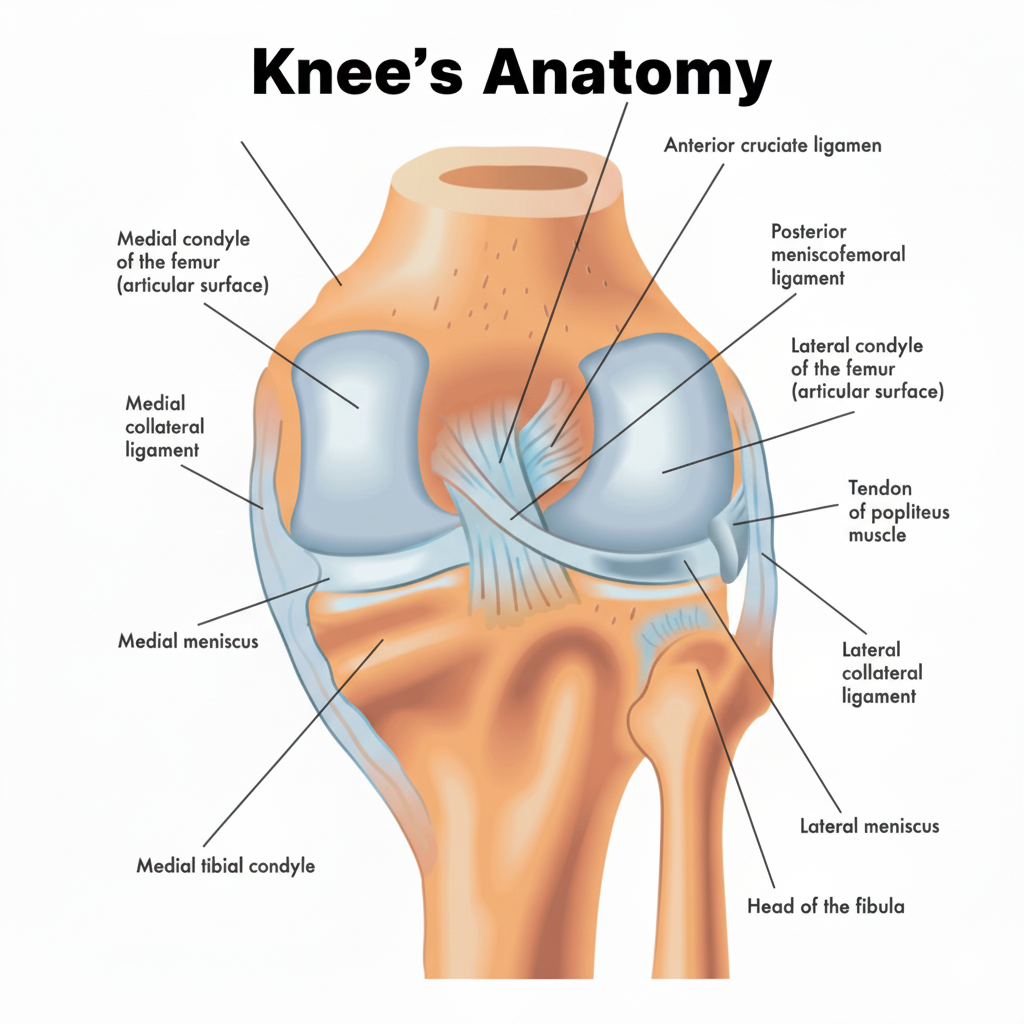
Ligaments
Think of ligaments as strong, slightly elastic ropes that connect your bones and provide stability. They prevent your knee from wobbling or moving in the wrong direction. There are four key ligaments:
Anterior Cruciate Ligament (ACL): Located in the center of the knee, it controls forward movement and rotation of the shinbone. ACL pain location is often described as a deep, internal pain.
Posterior Cruciate Ligament (PCL): Also in the center, it controls the backward movement of the shinbone.
Medial Collateral Ligament (MCL): Runs along the inner side of knee, providing stability against blows to the outer knee.
Lateral Collateral Ligament (LCL): Runs along the outer side, providing stability against blows to the inner knee. A ligament knee pain location chart helps distinguish which of these is injured.
Tendons
Tendons are tough, flexible cords that connect your muscles to your bones, allowing you to move your knee.
Quadriceps Tendon: Connects the powerful quadriceps muscles (front of your thigh) to your kneecap.
Patellar Tendon: Connects your kneecap to your shinbone. This is a common source of front knee pain.
Hamstring Tendons: Attach your hamstring muscles (back of your thigh) to the bones at the back of the knee.
Iliotibial (IT) Band: A thick band of tissue that runs from your hip down the outside of your thigh to your knee, crucial for stabilizing the knee during activities like running.
Cartilage and Meniscus
Articular Cartilage: A smooth, slippery substance that covers the ends of your bones, allowing them to glide effortlessly against each other. When this wears down (arthritis), you get knee bone on bone pain.
Meniscus: Two C-shaped pieces of tough, rubbery cartilage that act as shock absorbers between your femur and tibia. You have a medial (inner) and a lateral (outer) meniscus. A tear here is a very common cause of joint pain.
Now, let’s use this anatomical knowledge to explore the knee pain location chart.
Read – Pain in the Back of Knee When Straightening Leg After Sitting
The Knee Pain Location Chart: Mapping Your Discomfort
By identifying where your pain is concentrated, you and your doctor can narrow down the potential culprits. This knee pain diagram is a powerful first step in your knee pain diagnosis.
1. Pain in the Front of the Knee (Anterior Pain)
Pain felt at the front of the knee, around the kneecap, is extremely common. It often worsens when climbing stairs, squatting, or sitting for long periods.
Patellofemoral Pain Syndrome (Runner’s Knee): This is a general term for pain behind or around the kneecap. It’s often caused by overuse or misalignment of the patella, causing it to rub against the thighbone. The pain is a dull ache, and you might feel a grinding sensation.
Patellar Tendinitis (Jumper’s Knee): This is an inflammation of the patellar tendon, the structure connecting your kneecap to your shinbone. It causes pain below the knee cap and is common in athletes who jump frequently, like basketball or volleyball players.
Chondromalacia Patella: This involves the softening and breakdown of the articular cartilage under the kneecap. It leads to pain under kneecap and a grating feeling when you extend your knee.
Prepatellar Bursitis: The bursa is a small fluid-filled sac that reduces friction. Inflammation of the bursa in front of the kneecap (often from prolonged kneeling) causes swelling and tenderness right on the pain on knee cap.

2. Pain on the Inner Side of the Knee (Medial Pain)
The inner knee pain location chart points to issues with the structures on the inside part of your knee. This is a very frequent site of pain.
Medial Meniscus Tear: This is one of the most common knee injuries. A tear in the inner shock-absorbing cartilage can cause pain, swelling, stiffness, and a locking sensation. The pain on the inside of my knee is often sharp, especially when twisting. A twisted knee pain on inner side is a classic story for this injury.
MCL (Medial Collateral Ligament) Injury: An injury to the ligament on the medial side of knee often results from a direct blow to the outside of the knee. It causes pain and instability on the inner side.
Osteoarthritis: This “wear-and-tear” arthritis often affects the inner compartment of the knee more than the outer. It leads to a gradual onset of aching inner knee pain, stiffness (especially in the morning), and can be seen on a bad knee normal knee xray comparison.
Pes Anserine Bursitis: Inflammation of the bursa located on the lower inner side of the knee, causing tenderness and pain on inner side of knee.
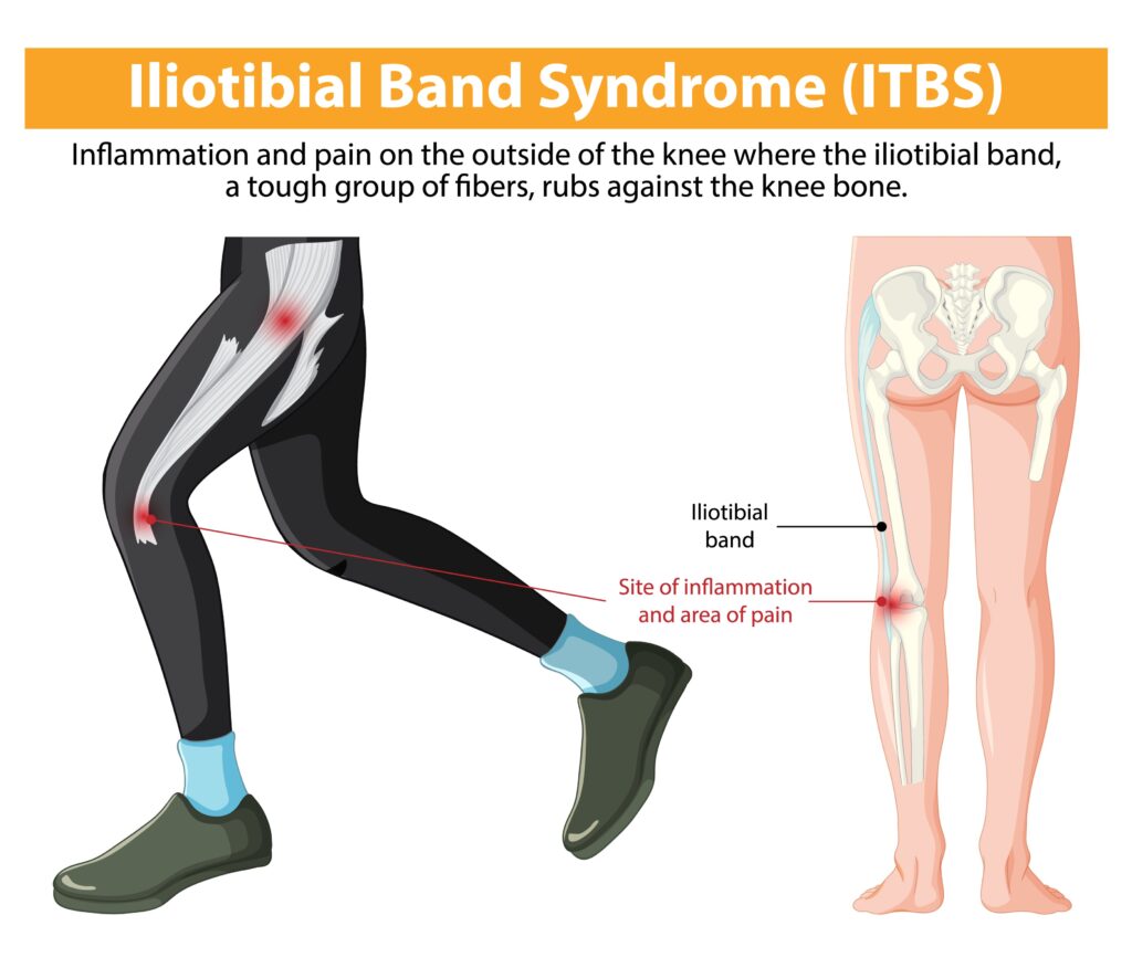
3. Pain on the Outer Side of the Knee (Lateral Pain)
Lateral knee pain is discomfort on the outside of your joint. This area is less commonly injured than the inner side but has specific causes.
Iliotibial (IT) Band Syndrome: A very common cause of outer side of knee pain, especially in runners and cyclists. The IT band rubs against the femur, causing inflammation and a sharp, burning pain on the outside of the knee. The pain on outside of knee when bending and straightening is a hallmark symptom.
Lateral Meniscus Tear: Similar to a medial tear, but occurring on the outer shock absorber. It causes knee side pain, swelling, and mechanical symptoms like catching or locking.
LCL (Lateral Collateral Ligament) Injury: An injury to the outer ligament, usually from a blow to the inside of the knee. It results in pain and instability on the outside.
Lateral Compartment Osteoarthritis: While less common than on the medial side, arthritis can also wear down the cartilage on the outer part of the knee joint.
4. Pain in the Back of the Knee (Posterior Pain)
The back knee pain location chart helps diagnose discomfort behind the joint, which can feel deep and hard to pinpoint.
Baker’s Cyst: This is a fluid-filled sac that forms a bulge at the back of the knee. It’s not usually a primary problem but rather a sign of another issue, like a meniscus tear or arthritis, causing the knee to produce excess fluid. It creates a feeling of tightness and pain behind knee that worsens when you fully extend or bend the knee.
Hamstring Tendinitis: Inflammation of the hamstring tendons where they attach to the back of the knee can cause a dull ache and stiffness. This tendon behind knee pain is common in athletes with tight hamstrings.
PCL (Posterior Cruciate Ligament) Injury: A tear in the PCL, often from a direct blow to the front of the knee (like a dashboard injury in a car accident), can cause deep back of knee pain and instability.
Gastrocnemius Tendinopathy (Calf Strain): An injury to the top of the calf muscle can also refer pain up to the back of the knee.
5. Pain Above the Knee
Discomfort felt directly above the knee often involves the large muscle group at the front of your thigh.
Quadriceps Tendonitis: Inflammation of the large tendon that connects the quadriceps muscles to the top of the kneecap. This causes pain right at the base of the thigh, just above the knee, especially when squatting, jumping, or climbing stairs.
Arthritis: In advanced cases, the inflammation and bone spurs from osteoarthritis can cause pain that radiates above the immediate joint line.
6. Pain Below the Knee
Pain felt on the shinbone just below the kneecap has a few distinct causes, often related to age and activity.
Osgood-Schlatter Disease: A common cause of knee pain in growing adolescents. It’s an inflammation of the area where the patellar tendon attaches to the tibia. It causes a painful lump and pain just below knee.
Patellar Tendinitis: As mentioned before, the pain from this condition is centered right on the tendon below knee cap above shin.
How a knee pain location chart helps
So, why does my knee hurt? By using this knee pain map, you’re no longer dealing with a vague, frustrating problem. You can start to connect the dots:
Improved Communication: Instead of just saying “my knee hurts,” you can tell your doctor, “I have a sharp pain on the inner side of my knee when I twist,” or “I feel a dull ache at the back of my knee that’s worse when I straighten it.” This specific information is incredibly valuable for a precise knee pain diagnosis.
Faster Diagnosis: Knowing the location helps your specialist focus their physical examination and choose the right imaging tests, like an X-ray (to check for arthritis or fractures) or an MRI (to look at soft tissues like ligaments and menisci). It helps differentiate between the many types of knee pain.
Informed Self-Care: While awaiting a professional diagnosis, understanding the likely source can guide initial self-care. For example, pain consistent with IT Band Syndrome might respond well to foam rolling the outer thigh, whereas pain suggestive of a meniscus tear requires rest and avoiding twisting motions.
However, self-diagnosis has its limits. This chart is a guide, not a substitute for professional medical advice.

From Rest to Replacement: A Spectrum of Treatment Options
Once you and your specialist have used the knee pain location chart to pinpoint a likely diagnosis, the next question is: what can be done about it? Treatment for knee pain is not one-size-fits-all. It exists on a spectrum, starting with simple, conservative measures and progressing to more advanced interventions only when necessary. An expert surgeon like Dr. Al-Khateeb will always explore the least invasive, most effective options first.
Tier 1: Conservative & At-Home Care
For many overuse injuries like mild tendinitis or strains, the first line of defense is the R.I.C.E. method:
Rest: Avoid activities that aggravate the pain. This gives the inflamed tissues time to heal.
Ice: Apply a cold pack for 15-20 minutes several times a day to reduce swelling and numb the pain.
Compression: Use an elastic bandage to help control swelling, but be careful not to wrap it too tightly.
Elevation: Keep your knee raised above the level of your heart as often as possible to help reduce swelling.
Tier 2: Physical Therapy & Medication
If home care isn’t enough, the next step often involves professional guidance:
Physical Therapy: This is a cornerstone of knee pain treatment. A physical therapist will design a program of specific exercises to strengthen the muscles that support your knee (like the quadriceps and hamstrings), improve flexibility, and correct any biomechanical issues that may be contributing to your pain.
Medications: Over-the-counter nonsteroidal anti-inflammatory drugs (NSAIDs) like ibuprofen can help manage pain and inflammation. However, it is essential to consult with your doctor before starting any medication.
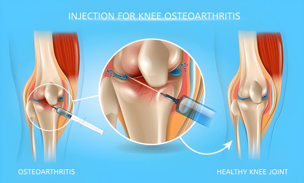
Tier 3: Injections & Minimally Invasive Procedures
For more persistent pain, particularly from conditions like arthritis or severe bursitis, your doctor may suggest:
Corticosteroid Injections: A powerful anti-inflammatory medication injected directly into the knee joint can provide rapid, though temporary, pain relief.
Viscosupplementation: Injections of hyaluronic acid, a substance that mimics natural joint fluid, can help lubricate the knee joint and reduce the pain of osteoarthritis.
Tier 4: Surgical Intervention
When conservative treatments fail to provide relief, or in cases of severe structural damage (like a complete ACL tear or advanced arthritis), surgery may be the best option to restore function and eliminate pain.
Arthroscopy: A minimally invasive “keyhole” surgery where a surgeon can repair meniscus tears, reconstruct ligaments like the ACL, or remove loose pieces of cartilage.
Knee Replacement (Arthroplasty): For end-stage arthritis where the cartilage is gone (knee bone on bone), replacing the damaged joint surfaces with artificial components can be a life-changing procedure. This is a highly specialized field where the experience of the surgeon, like Dr. Al-Khateeb’s extensive record of over 2,000 replacements, is paramount.
Consult Dr. Hesham Al-Khateeb: Expert solutions with a knee pain location chart
While minor aches and pains can often be managed at home, certain symptoms warrant an immediate consultation with an expert knee surgeon. As a leading specialist in Dubai, Dr. Hesham Al-Khateeb advises patients to seek professional help if they experience:
Inability to bear weight on the knee.
Severe swelling, pain, or obvious deformity.
A “pop” at the time of injury.
A knee that locks, catches, or feels unstable.
Pain that doesn’t improve after a few days of self-care.
Fever, redness, and significant warmth, which could indicate an infection.
Ignoring persistent knee pain can lead to further damage and more complex problems down the line.
Dr. Hesham Al-Khateeb is an Award-Winning UK Board-Certified Hip and Knee Surgeon with over 2,000 joint replacements performed. Trained across London, Canada, Hamburg, and Seattle, he brings a global perspective to his Dubai practice. His expertise covers hip & knee replacements, revision surgery, hip arthroscopy, ACL & meniscal repair, sports injuries, and trauma. Recipient of the NHS Innovation Award and Norman Capener Award, Dr. Al-Khateeb is recognized for advancing orthopedic care and delivering personalized treatment at the highest standards.
Your knees are engineered for a lifetime of movement. Don’t let pain stop you from living your life to the fullest. By using the knee pain location chart to understand your symptoms and seeking timely expert care, you can regain your mobility and get back to the activities you love.
Don’t let knee pain dictate your life. Combat it and regain your mobility – get in touch with Dr. Hesham Al-Khateeb’s team today!
Frequently Asked Questions (FAQs)
The most common types of knee pain stem from either acute injury (like a meniscus tear or ACL sprain), overuse conditions (like tendinitis or IT band syndrome), or degenerative conditions like osteoarthritis. The specific type greatly depends on a person’s age, activity level, and medical history. Using a knee pain chart can help narrow down the possibilities based on the pain’s location.
While the causes of knee pain are similar, some conditions are more prevalent in women. For example, women are more likely to suffer ACL injuries and develop Patellofemoral Pain Syndrome due to anatomical differences in pelvic width and leg alignment (a wider “Q-angle”). Osteoarthritis is also more common and can be more severe in women after menopause. Understanding the specific factors related to knee pain in ladies is crucial for proper diagnosis and treatment.
A normal knee xray will show smooth bone surfaces and a clear, even space between the femur and tibia, which represents healthy cartilage (cartilage itself is not visible on X-ray). A bad knee xray, particularly one with osteoarthritis, will show a narrowed joint space, indicating cartilage loss. You might also see bone spurs (osteophytes) and changes in the bone’s density, indicating a knee bone on bone condition.
The most common causes for pain on the inner side of the knee are a tear of the medial meniscus, a sprain of the MCL (medial collateral ligament), or medial compartment osteoarthritis. An acute, twisting injury is more likely to cause a meniscus or MCL tear, while a gradually worsening ache is more typical of arthritis. An expert can make an accurate knee pain diagnosis after a physical exam and appropriate imaging.
A sharp pain behind knee could be caused by several issues highlighted on the back knee pain location chart. A Baker’s Cyst can cause a feeling of intense pressure and sharpness. A tear in the root of the meniscus or an injury to the hamstring tendons can also cause sharp posterior pain. It’s important to get this evaluated, as it could signal a significant underlying issue within the joint.